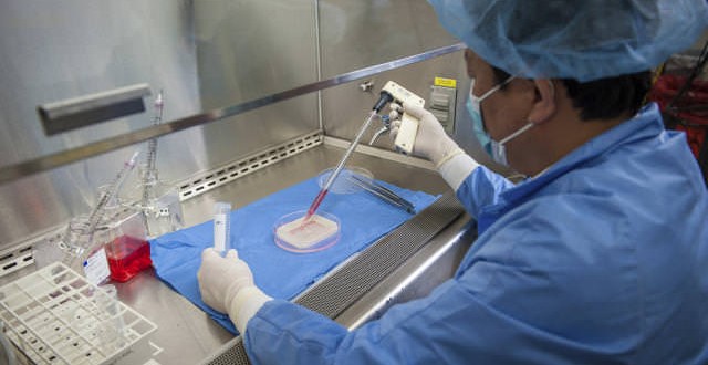
For the first time, researchers have successfully implanted laboratory-grown vaginas in human patients—potentially helping women avoid drawbacks from other regenerative procedures, scientists announced in a paper published today.
A research team at Wake Forest Baptist Medical Center’s Institute for Regenerative Medicine, led by Dr. Anthony Atala, said in Lancet that four recipients of lab-grown reproductive organs underwent surgery between June 2005 and October 2008—and that the organs are functioning normally after eight years. The new organs are created by culturing these women’s own cells into tissues and eventually reshaping the tissues into a vagina-like structure.
All four of the women in the study were born with Mayer-Rokitansky-Küster-Hauser (MRKH) syndrome, a rare genetic condition in which the vagina and uterus are underdeveloped or absent.
Conventional treatment generally involves the use of grafts made from intestinal tissue or from skin, but both tissues have drawbacks, says Dr Atala, a paediatric urologic surgeon at Wake Forest.
Intestinal tissue produces an excess of mucus, which can cause problems with odour. Conventional skin, meanwhile, can collapse.
Dr Atala said women with this condition usually seek treatment as teenagers.
“They can’t menstruate, especially when they have a severe defect where they don’t have an opening,” he said.
This can cause abdominal pain as menstrual blood collects in the abdomen. “It has nowhere else to go,” he said.
Girls in the study were aged 13 and 18 at the time of the surgeries, which were performed between June 2005 and October 2008.
The researchers started off by collecting a small amount of cells from genital tissue and grew two types of cells in the lab: muscle cells and epithelial cells, a type of cell that lines body cavities.
About four weeks later, the team started applying layers of the cells onto a scaffold made of collagen, a material that can be absorbed by the body.
They then shaped the organ to fit each patient’s anatomy, and placed it in an incubator.
A week later, the team created a cavity in the body and surgically attached the vaginal implants to existing reproductive organs.
Once implanted, nerves and blood vessels formed to feed the new organ, and new cells eventually replaced the scaffolding as it was absorbed by the body.
“By the six-month time point, you couldn’t tell the difference between engineered organ and the normal organ,” Dr Atala said.
The team continued to monitor the young women, taking tissue biopsies, MRI scans and internal exams, for up to eight years from the initial implants.
All of these tests showed the engineered vaginas “were similar in make up and function to native tissue”, said Atlantida-Raya Rivera, director of the HIMFG Tissue Engineering Laboratory at the Metropolitan Autonomous University in Mexico City, where the surgeries were performed.
Professor Martin Birchall of UCL Ear Institute in London, who wrote a commentary in the same journal, said the findings address some important questions about tissue-engineering, including whether tissue will grow as patients grow and whether an organ as large as the vagina can develop blood vessels when implanted in the body.
Agencies/Canadajournal
 Canada Journal – News of the World Articles and videos to bring you the biggest Canadian news stories from across the country every day


