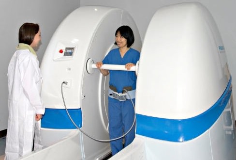
The University of B.C. says it’s making progress using a state-of-the-art MRI machine – of which only 12 exist in the world – to study joint aches.
The machine is an upright, open MRI that allows a subject to sit, stand or squat while having the procedure, so the effects of the activities on a person’s joints can be observed in greater detail.
“This machine tells us a lot more about people’s joints than normal MRI,” says biomedical engineer David Wilson, a professor in UBC’s Department of Orthopaedics and researcher at the Vancouver Coastal Health Research Institute’s Centre for Hip Health and Mobility.
“With normal MRI all we get is a snapshot of the damage to the tissues in the joint. What this scanner lets us do is scan the joints in action so we can gain much greater insight into what’s causing the pain and tissue degeneration,” Wilson says.
Joint effort
Being able to capture joints in different positions is essential to Wilson and his team in their quest to learn more about joint pain.
They’re particularly interested in studying hip osteoarthritis, a painful condition caused by the wear and tear of the hip joint, which allows us to sit, stand, run and walk.
Understanding hip osteoarthritis–which affects up to one million Canadians, many of whom are seniors–is of critical importance. Since there is no cure early prevention is key, which is why Wilson and his team are following a group of middle-aged people over the next five years to monitor their hip health.
“We want to learn more about people who are at risk before they get the disease,” Wilson explains. “We want to find out why some people have their joints degenerate very quickly, and why some have their joints degenerate slowly.”
One of a kind
Currently, the machine at VGH is the only one in the world solely used for research purposes. Scans from the machine can reveal rare views of virtually every part of the body from the lungs to the spine to the feet. Blood flow and liquids can also be captured, including the build-up of fluid around the joints.
While the scanner has the potential to be used clinically, especially for imaging the body in positions that cannot be captured in the confines of traditional MRI, more research is needed before standing up for a scan becomes the new normal.
In the future, Wilson’s team hopes to establish definitive guidelines and protocols doctors can follow to advise individual patients on the best treatment option for their pain, whether it be medication, physical therapy or hip replacement surgery.
“If we can learn more about the cause of joint pain by leveraging the unique capabilities of this machine, we’re in a much better position to design effective treatment and prevention strategies for osteoarthritis,” Wilson says.
Agencies/Canadajournal

 Canada Journal – News of the World Articles and videos to bring you the biggest Canadian news stories from across the country every day

